structural-biology/ai produced the result/Frontiers in Molecular Biosciences 2022 · v2
Researchers read an AlphaFold model of honey bee vitellogenin's tail end
A study examined a computer-predicted structure of the honey bee egg-yolk protein vitellogenin, made with the AlphaFold neural network, and proposed that its tail region swings over to cover the protein's fatty cargo pocket.
spectrum · one line per step, placed by what the step does · bright lines used AI
How Honey Bee Vitellogenin Holds Lipid Cargo: A Role for the C-Terminal
Frontiers in Molecular Biosciences, 2022
doi:10.3389/fmolb.2022.865194 · record aix-00195 v2 · checked 2026-10-09
- AI was for
- Structure determination
- Model family
- Transformer
- Checked by
- None stated
- Code
- not reported
The finding the paper is about came from the AI.
What this research was about
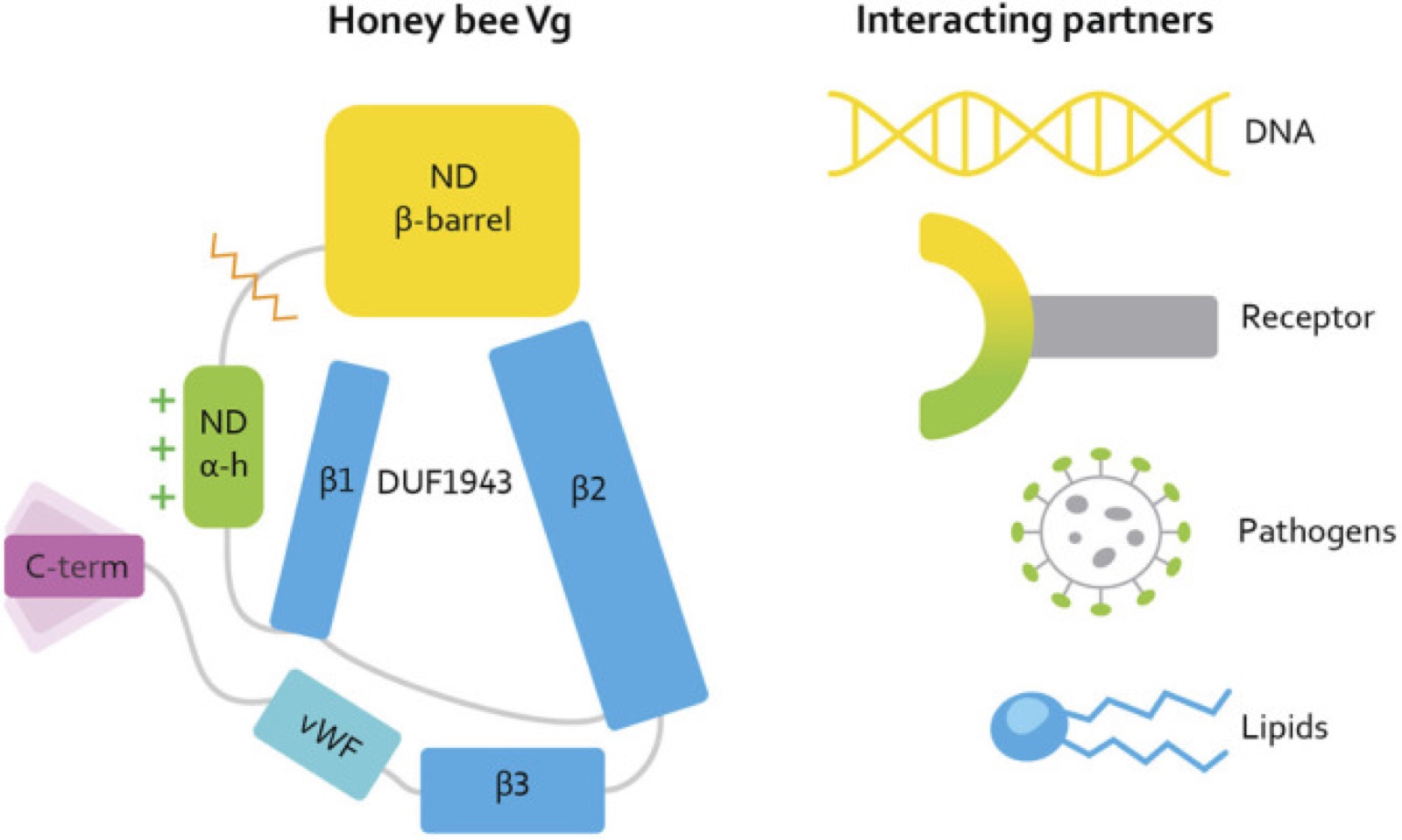
Vitellogenin is a large protein that insects, worms, fish and many other animals use to carry fat and nutrients to their eggs. In honey bees it has other jobs too. Carrying fat is awkward work: fatty molecules do not mix with water, so the protein has to hold them inside a greasy pocket, away from the watery surroundings of the body. Working out how it does that means knowing the protein's three-dimensional shape, and shapes of very large proteins are hard to measure. The usual methods, such as growing crystals or imaging frozen samples, often fail or give only a blurred outline.
The authors had earlier produced a predicted shape for the whole honey bee protein and fitted it to a low-resolution electron microscopy map, a fuzzy picture showing roughly where the protein's bulk sits. That map suggested there was space above the fatty pocket. Here they look closely at one part of the predicted structure, the C-terminal region, meaning the tail end of the protein chain, and ask what it is for.
Where AI came in
The shape itself came from AlphaFold, a neural network from DeepMind that predicts how a protein chain folds up from the sequence of its building blocks. It was used off the shelf, with no training or tuning in this study, and the honey bee prediction was generated in the authors' preceding paper and re-analysed here. The researchers also took AlphaFold's database prediction for the equivalent protein in the nematode worm C. elegans, alongside two experimentally solved structures from the public Protein Data Bank, for comparison.
In effect AlphaFold stood in for a measurement nobody has made: there is no experimentally solved structure of honey bee vitellogenin in the paper, so every structural claim rests on the predictions. Everything after the prediction was done by the researchers with standard software and their own reading. They overlaid the honey bee and worm tail regions, giving a close match, mapped surface electrical charges, counted the chemical cross-links holding each fold together, and from that proposed the open-and-shut shielding mechanism.
Written by AIxSci from the checked record below, to give context for readers outside the field. It is not part of the record.
The work
Technical · from the record
The authors analyse an AlphaFold prediction of full-length honey bee vitellogenin, generated in their earlier work, to ask what its folded C-terminal region does. Superimposing the predicted honey bee C-terminal region (residues 1688-1770) on the AlphaFold database prediction of the C. elegans Vg-2 C-terminal region (residues 1530-1613) gave an RMSD of 1.035 with an almost identical fold, while the honey bee fold has three disulfide bridges and Vg-2 has two. Combining this with surface-charge maps, disulfide positions and an insect-specific loop seen in the model, the article proposes that a flexible linker lets the C-terminal region move between a position flanking the protein and one covering the opening of the hydrophobic lipid binding cavity.
How AI was used
AlphaFold was used off the shelf to predict the full-length structure of honey bee vitellogenin from its sequence; that prediction was produced in the authors' preceding publication and is re-analysed here, together with the model's earlier fitting into a low-resolution EM map that indicated available density above the lipid binding site. The authors also took the AlphaFold database prediction of C. elegans Vg-2 and the experimental structures PDB 1LSH and 6I7S as comparison structures. On these models they performed structural superposition of the C-terminal regions with RMSD calculation, electrostatic surface calculation with the APBS plugin in PyMol, inventories of disulfide bridges and hydrophobic and electrostatic contacts, sequence alignment, and comparison of connecting alpha-helix lengths across species. No model was trained or fine-tuned in this study, and the proposed conformational mechanism was derived by expert reading of these predicted structures rather than by computation.
The shape of the work
Structural · the record, drawn
AI
Predict full-length honey bee Vg structure
Running a trained model over new data to predict, classify or score. The AI stood in for unresolved measurement.
Using AlphaFold and EM contour mapping, we recently described the protein structure of honey bee Vg.where the paper describes this · verbatim
no AI
Fit prediction into low-resolution EM map
Testing outputs against ground truth.
Our previous study fitted the AlphaFold prediction into a low-resolution EM map.where the paper describes this · verbatim
no AI
Retrieve homologous structures for comparison
Obtaining raw data, whether by measurement, download or retrieval.
The datasets analyzed for this study can be found in the PDB at https://www.rcsb.org/ (PDB-ID: 1LSH and 6I7S)where the paper describes this · verbatim
no AI
Superimpose C-terminal folds across species
Extracting understanding from model behaviour.
Superimposing the C-terminal region (amino acid 1530–1613) in C. elegans Vg-2 with our prediction of the C-terminal in honey bee Vgwhere the paper describes this · verbatim
no AI
Map charges, disulfide bridges and contacts on the model
Extracting understanding from model behaviour.
the electrostatic charges are calculated using the APBS plugin in PyMolwhere the paper describes this · verbatim
no AI
Propose open/closed C-terminal shielding mechanism
Extracting understanding from model behaviour.
We propose that the C-terminal region provides this shielding.where the paper describes this · verbatim
What the record says
Technical · every part carries its own basis
+ in the paper~ our reading− not reported
How to read the quotations. A quotation shows where the paper describes something. It does not quote every value beside it: one passage locates a part of the work, and values without their own quotation are our reading of that passage.
Every structural claim in the article rests on AlphaFold predictions: the authors' full-length honey bee Vg model (generated in their prior work and analysed here) and the AlphaFold database model of C. elegans Vg-2. No experimentally solved structure of honey bee Vg exists in the paper.
Recent progress made possible by DeepMind’s AlphaFold, a neural network for structure predictionwhere the paper describes this · verbatim
Publicly available datasets were analyzed in this study.where the paper describes this · verbatim
What this paper did not report
Technical · absence is published deliberately
- CodeWhether the code is available is not stated.
- Trained model weightsWhether the trained model is available is not stated.
- ComputeThe hardware or time used is not stated.
- ValidationNo validation of the AI is described.
- Version of AlphaFoldWhich version of the model was used is not stated.
About this article
Record aix-00195, version 2, checked by a person on 2026-10-09. The record describes the paper; it does not assess whether the paper's findings are right. The paper is published under CC-BY; quotations are at most 25 words. How we work · Report an error